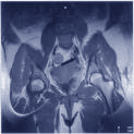
Fat-suppressed MRI
images show high signal intensity of vaginal melanoma
(Malignant
Melanoma August 28, 2003)
"We report two cases of
vaginal melanoma with magnetic resonance imaging findings," scientists in
South Korea report.
"The first melanoma was
a bilobular polypoid mass with melanotic and amelanotic components, which
arose from the lateral wall of the vaginal canal. The melanotic melanoma
showed high signal intensity (SI) on T1-weighted images and low SI on
T2-weighted images, which was not suppressed by a fat-saturated sequence,"
wrote H. Kim and colleagues, Catholic University of Korea, Department of
Radiology.
The researchers
concluded: "Another melanoma showed a pale brown polypoid mass in the
vagina, which revealed intermediate SI on T1-weighted images and high SI on
T2-weighted images. On fat-suppressed images, both tumors were more clearly
demonstrated with high SI."
Kim and colleagues
published their study in the Journal of Computer Assisted Tomography
(Magnetic resonance imaging of vaginal malignant melanoma. J Comput Assist Tomogr, 2003;27(3):357-360).
For more information,
contact H. Kim, Catholic University of Korea, Department of Radiology,
Daejeon St. Mary's Hospital, 520-2 Daeheung Dong, Taejon 301723, South
Korea.
The information in this
article comes under the major subject areas of Medical Devices and
Gynecology. This article was prepared by Women's Health Weekly editors from
staff and other reports.
ęCopyright 2003, Women's
Health Weekly via NewsRx.com & NewsRx.net