Keratoconus: causes, symptoms and treatment

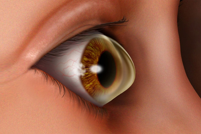
Keratoconus is a progressive eye disease in which the normally round cornea thins and begins to bulge into a cone-like shape. This cone shape deflects light as it enters the eye on its way to the light-sensitive retina, causing distorted vision. Keratoconus can occur in one or both eye's.
Symptoms of Keratoconus
In its earliest stages, the symptoms of keratoconus are slight blurring, distortion of vision, nearsightedness, astigmatism, and increased sensitivity to glare and light. In rare cases, a sudden, significant decrease in vision will occur. The sudden and significant decrease in vision is caused by swelling of the the cornea. The swelling may last for weeks or months.

What Causes conus?
The weakening of the corneal tissue that leads to keratoconus may be due to an imbalance of enzymes within the cornea. This imbalance makes the cornea more susceptible to oxidative damage from compounds called free radicals, causing it to weaken and bulge forward.
Risk factors for oxidative damage and weakening of the cornea include a genetic predisposition, explaining why keratoconus often affects more than one member of the same family.
Keratoconus is also associated with
- overexposure to ultraviolet rays from the sun,
- excessive eye rubbing,
- a history of poorly fit contact lenses, and,
- chronic eye irritation.

Treatment Options
The treatment approach to keratoconus follows an orderly progression from
glasses to contact lenses to corneal transplantation. Glasses are an effective
means of correcting mild keratoconus. As the cornea steepens and becomes more
irregular, glasses are no longer capable of providing adequate visual
improvement. Corneal transplant surgery is indicated when a patient cannot wear
contact lenses (corneal or scleral) for an acceptable period of time or when the vision, even with
contacts, is unsatisfactory. Over 90% of corneal transplants are successful
with the majority of patients obtaining vision of 20/40 or better afterwards
with either glasses or contact lenses.
A gas permeable contact lens is the most highly effective way to manage keratoconus and 90% of all cases can be managed this way indefinitely. If the cornea becomes too scarred or painful, a corneal transplant may be necessary.
When is it necessary to face a cornea transplant?
When there is a severe thinning of the cone's summit or when central dull scars interfere with vision, , in order to improve their vision and in order to end pain caused by contact lenses, patients must undergo a cornea transplant.
Deep Anterior Lamellar Keratoplasty (DALK)
With advancement in corneal surgical techniques, it is now possible to
selectively remove the anterior layers from the cornea and replace it with
donor tissue to restore its
anatomy and
function. Deep anterior lamellar keratoplasty (DALK) is one such
procedure wherein the host corneal endothelium is retained, and anterior
corneal tissue is replaced with normal thickness donor tissue. As the host
endothelium is retained there is no risk of rejection, and steroids
have to be given only for a short duration of time. However DALK surgery
requires more surgical expertise compared to the traditional full thickness
keratoplasty, and hence performed by only well trained corneal surgeons all
over the world.
In advanced stages of Keratoconus, due to extreme
thinning, the inner layer of the cornea can rupture, leading to increased
leakage of fluid into the cornea. This results in whitening of the cornea,
with sudden decrease in vision. This condition is called acute hydrops.
In this situation, topical medications have to be applied for symptomatic
relief. It takes 3 - 4 months for the corneal oedema to resolve, following
which a standard full thickness keratoplasty is required to restore corneal
clarity and visual improvement. It this situation lamellar surgery is not
recommended, and hence one should not wait for this complication to occur,
and take advantage of lamellar procedures at an early stage.
Standard full thickness corneal transplantation procedure, is very successful in restoring corneal structure and function. However one needs to use topical steroids for a longer duration than lamellar procedures. Full thickness corneal grafts in keratoconus have the best clinical outcome, when compared with other indication of corneal transplantation surgery.
In addition to treatment, the following tips may be helpful:
-
Wear UV protected
sunglasses while outdoors to reduce your exposure to
ultraviolet light.
Avoid vigorous eye rubbing. Rubbing the eye's may increase the effects of keratoconus.
Consult an allergist to manage allergies appropriately. Recommendations may include environmental changes or prescription allergy medications.
See your eye doctor on a regular basis. Ensure that contact lenses are properly fitted to produce the clearest vision.
Use preservative-free artificial tears and lens care products. These products are compatible with the eye�s surface.
