

fitness news
![]()
![]()
Font size Women’s Health

New Way to Look Inside the Body
– Reported, November 26, 2012
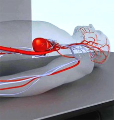

WILMINGTON, DE (Ivanhoe Newswire) – Its being called the biggest advance in imaging for the spine since the invention of x-rays in 1890.
Kate Gordon has high hopes for a career in sports.
“Play soccer for the Olympics, yeah in that category,” Kate told Ivanhoe.
Then she found out she had scoliosis.
“Like do I need surgery? What will happen in the future?” Kate said.
Kids like Kate need to get two x-rays a year to monitor their spines until they stop growing. Her mom was concerned about excessive radiation.
“Especially since she hadnt entered her growth spurt yet, they were wanting to follow it closely,” Anne Gordon, Kates mom, said.
Thats where this technology comes in.
“Its the biggest development in x-rays since the invention of x-rays,” Dr. Suken A. Shah from Nemours Alfred I. Dupont Hospital for Children in Wilmington, DE told Ivanhoe.
The EOS) imaging system takes a full body scan in minutes and emits up t0 90 percent less radiation than a traditional CT scan.
“It might prevent their chance of having a malignancy as an adult,” Dr. Shah said.
The machine gives physicians the ability to study Kates spine in 3-D, allowing them to better treat patients.
“The clarity and the detail is much, much better than we were ever able to see on conventional x-rays. You can see the 3-dimensional deformity where theres a rib hump on this side and theres quite a twist up to the spine and weve been able to eliminate a lot of that with this new instrumentation,” Dr. Shah said.
“Its a much better thing for them and its a huge relief for a parent as well,” Anne Gordon said.
And its helping keep Kates sports dreams alive.
The Nobel Prize winning technology isnt just used to scan the spine. It can also be used for other parts of the body.