Deep-vein thrombosis is the formation of blood clots in veins deep inside the legs. The condition is usually caused by sluggish blood flow when a person lies or sits still for long periods of time, such as during prolonged bed rest after surgery or in cases of paralysis. Prevention of thrombosis is one of the reasons your are told to get up and walk around as soon as possible after having an operation.
Deep-vein thrombosis is more common among women over 35 who smoke and take birth-control pills, or women who are or recently have been pregnant. Deep-vein thrombosis is not always a serious condition but, if a piece of a blood clot breaks off and travels to your lungs, it can block an artery, which can be life threatening. Deep venous thrombosis (DVT) affects mainly the veins in the lower leg and the thigh.
Risk Factors associated with DVT:
Risks include
- Prolonged sitting,
- Bed rest or immobilization (such as on long plane or car trips),
- Recent surgery or trauma (especially hip, knee or gynecological surgery),
- Fractures,
- Childbirth within the last 6 months and the use of medications such as estrogen and birth control pills.
- Coronary heart disease
- Being overweight or obese
Risks also include
- a history of polycythemia vera
- malignant tumor, and
- inherited or acquired hypercoagulability.
Symptoms
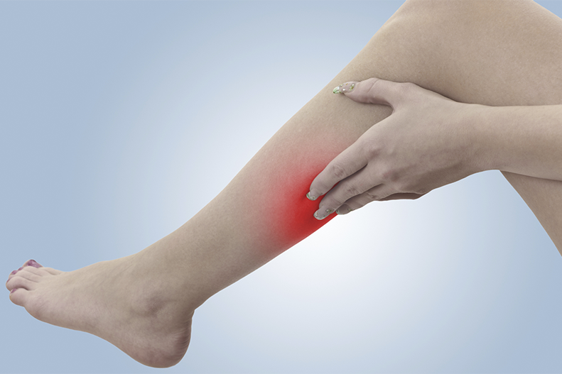
A clot in a vein in your leg can cause a variety of symptoms, including pain, tenderness, swelling, redness, and a feeling of warmth on the skin over the clot. Specific symptoms include:
- leg pain in one leg only
- leg tenderness in one leg only
- swelling (edema) of only one leg
- increased warmth of one leg
- changes in skin color of one leg, redness
Diagnosis
Deep-vein thrombosis can be diagnosed by
- Radionuclide scanning – In a radionuclide scan, the tracer either is injected into a vein or swallowed. Once the tracer enters the body, it travels through the bloodstream to a target organ, such as the thyroid, heart or bones. Different tracers tend to collect in different organs. The tracer emits gamma rays, which are similar to X-rays. These gamma rays are detected by a gamma camera and analyzed by a computer to form an image of the target organ. Sites of potential problems emit more intense gamma rays and appear as bright spots on the scan. PET scans, gallium scans and bone scans are all types of radionuclide scans.

- An ultrasound – DVTs are most commonly detected nowadays by use of ultrasound – scientists say they can now use this method to detect even the smallest of clots. A water-soluble gel is placed on the transducerand the skin over the veins of the extremity being tested. To examine the arteries: Blood pressure cuffs will be put around the thigh, calf, and ankle to examine the legs. In the arms, the blood pressure cuffs are placed at different points along the arm. A conductive paste is applied to the skin over the arteries being examined. The cuff will be inflated above the normal systolic blood pressure for the extremity. The transducer is placed near the cuff, and the pressure in the cuff is released slowly. When the “swishing” is detected, it is recorded as the blood pressure. This is repeated for each cuff.
- D-dimer blood test
- venography of the legs
These imaging and blood tests can provide information about the condition of the veins in your legs and the flow of blood through them.
Treatment Options
- If the blood clots are small and confined to your calf, you may not need any treatment.
The clots may break up and dissolve by themselves, especially if you walk around frequently.
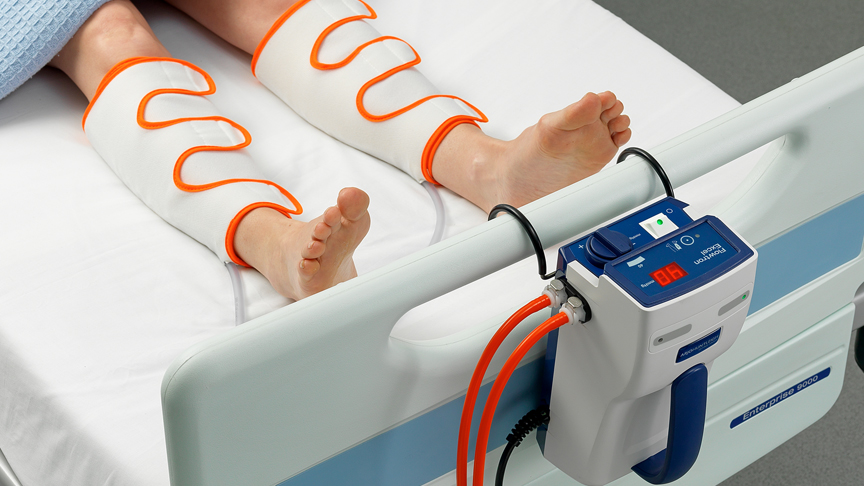
Too much bed rest is discouraged . - Medical compression stockings are often worn to cover the length of the whole leg to give support to the veins and reduce swelling.
- If you have a serious case of thrombosis, your doctor may prescribe drugs that thin your blood and prevent clotting, especially if there is a risk of a pulmonary embolism. This usually initially involves giving high doses of the drug heparin by injection. Patients are also prescribed a similar drug, warfarin, in tablet form, which they may stay on for several months. When taking these blood thinning drugs patients usually have regular blood tests to make sure they are getting the right dose and are not at risk of a haemorrhage. Clots that are located in the deep veins in the thigh or behind the knee are more likely to break off and travel to the lungs. It is important to take the medication exactly as directed by your doctor or pharmacist to ensure that you are not at risk to develop another DVT or increased bleeding.
- Surgery to remove a clot is sometimes necessary.
- Reducing risk factors such as quitting cigarettes, losing excess body fat and switching to a low fat diet.
Prevention
Anticoagulants may be prescribed as a preventive measure for high risk people or people undergoing high risk surgical procedures. Minimize immobility of the legs. In order to reduce the risk of DVT include treatment, such as reducing excess body fat, quitting cigarettes, exercising regularly and switching to a high fibre, low fat diet.
- Exercise the legs regularly – take a brisk 30-minute walk every day. Lower extremity exercises such as simple leg lifts, elevating the foot of the bed, and active and passive ankle motion to increase blood flow through the femoral vein.
- Maintain a weight that’s appropriate for your height
- Avoid sitting or lying in bed for long periods of time without moving the legs
- Women, particularly those over the age of 35, should consider the risks and benefits of taking the contraceptive pill
Disclaimer
The Content is not intended to be a substitute for professional medical advice, diagnosis, or treatment. Always seek the advice of your physician or other qualified health provider with any questions you may have regarding a medical condition.